Health Benefits and Toxicity of the Element Tin and its Effect on Adrenals, Depression and Fatigue
While Tin (Sn) has been established to be an essential trace element for some animals (they won't grow well without it), some researchers are still unsure of whether tin is essential in human health and nutrition. Daily dietary intake of tin from various food sources is in the 1-3 mg range, which is less than 1/10th of the daily intake obtained years ago before lacquering tin cans, switching to aluminum cans, or in the more distant past, when tin cups or tin pans were still in use. Since bronze contains copper and tin, the use of tin has been established well past the Bronze Age, several thousand years ago.
Rat studies have shown that tin-deficient diets resulted in poor growth, reduced feeding efficiency, hearing loss, and bilateral (male pattern) hair loss. Tipton and Shafer examined tin in human tissue after accidental deaths. They noted that tin was found in the aorta, heart, kidney, liver, muscle, ovary, spleen, pancreas, brain, testes, stomach, and uterus, but none was found in the thyroid of any victim, while the prostate, which usually shows no other trace element, had tin.
Average concentrations were the same range as cobalt, chromium, iodine, and selenium, which are known vital nutrients. Inorganic tin is capable of entering into biological activity at saline pH, and it is far less toxic than other known vital trace elements such as copper and cobalt. In addition, tin levels do not vary statistically with gender, age, or geographical areas. Misk found traces of tin in the fetal heart and spleen, and higher levels in the liver, while Schroeder and others reported no tin in stillborns.
Tin is associated with Iodine the same way as calcium is associated with magnesium (see "Tin & Iodine" for details). Tin supports the adrenal glands, and iodine supports the thyroid gland, with both subsequently affecting cardiac output: Tin + adrenals control the left side, and iodine + thyroid control the right side. In addition to low Vitamin C and/or Vitamin B1, low tin is a common nutritional cause of low adrenals, which can lead to left-sided cardiac insufficiency. While fatigue or depression may be experienced with cardiac insufficiency of either side, breathing difficulties or asthma are more common with left-sided cardiac insufficiency, and swelling of hands and feet is more common with right-sided cardiac insufficiency, regardless of the cause.
Comparing thousands of patient records showed that better than 90% of patients tested exhibited moderately low, to very low levels of Tin when referenced to the status of all other essential trace minerals, making tin the most deficient element compared to any other trace mineral measured.
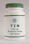
Dr. Ronald Roth had 285 individuals taking part in the Nutritional evaluation of Tin, some on a short-term basis (3 weeks), and others on a long-term basis (1 - 2+ years), resulting in some valuable feedback on various responses encountered, including side effects, although the rather poor absorption of stannous oxide was a limiting factor in being able to achieve optimal cellular levels of tin in all subjects.
Of the changes experienced after supplementing tin, negative reactions, e.g. stomach / digestive upsets, or skin reactions, were at par or less compared to the best tolerated trace minerals such as chromium, calcium, or magnesium. Positive health effects were numerous and included improvements with fatigue, some forms of depression, and a general increase in energy, well-being, and mood. There were also benefits with certain types of headaches, insomnia, asthma, or improvements with digestion, skin, or various aches and pains.
Tin toxicity[1] - or its health hazards - documented over the last 200 years in humans has been linked to the consumption of foods or beverages that were stored in tinned, unlacquered containers under long-term, low pH conditions, and where levels of several hundred to several thousand mg/kg were ingested. Symptoms were limited to mostly gastrointestinal complaints such as nausea, abdominal pain and vomiting, with excess tin being rapidly excreted, and no long-term negative health or toxic effects reported.
There are many causes of depression, some resulting from abnormal brain chemistry, while others are associated with low blood pressure, low thyroid, or low (or high) levels of various essential nutrients such as lithium, calcium, magnesium, copper, sodium, protein, Vitamin B1, B6, B12, manganese (low blood sugar), and others. Many of these nutrients are well documented in affecting mood, but Dr. Ronald Roth had not previously come across any reference to tin until starting to do research on it, and after it helped some patients with depression where any other drug, nutrient, or intervention had failed.
Tin is not a panacea for depression --- it will not work when other chemical imbalances are involved, but it can be the missing link when most other attempts to resolve depression have failed; essentially involving low, or malfunctioning adrenals. For the same reason, some cases of asthma - particularly when related to low adrenals and subsequent left-sided cardiac insufficiency - respond to tin as well.
Tin (as stannous fluoride) is found in some toothpastes, and it has been used in the form of stannous chloride as a chemical preservative. It is also added to asparagus to improve its taste, while in some countries it has been utilized as a remedy for intestinal parasites. Herbal Sources of tin (in the highest to lowest order) include doggrass, juniper, bilberry, milk thistle, dulse, lady slipper, althea, valerian, Irish moss, nettle, barberry, yarrow, blessed thistle, red clover, yellow dock, kelp, licorice, devils claw, pennyroyal, and senna.
Evidence Review
Critical Scientific Evaluation of Tin in Human Nutrition and Physiology
A Comparative Review of Historical Alternative Claims and Contemporary Biomedical Evidence
AI-assisted literature review ·
26 peer-reviewed sources ·
The article above is preserved as first published and has not been altered. What follows
is a separate document that examines its claims against the current biomedical
literature, reproduced here in full. The first section addresses the paired
tin–iodine model shared with the companion
Tin & Iodine article; the sections after it address tin
specifically.
The Paired Tin–Iodine Model
Both the tin and the tin/iodine articles on this site rest on a shared organising idea. Tin is
described as supporting the adrenal glands and iodine as supporting the thyroid gland, the two
elements are said to pair with one another in the way calcium pairs with magnesium, and the
pairing is then mapped onto the heart: tin and the adrenals are held to govern
left-sided cardiac output, iodine and the thyroid right-sided cardiac output.
From that mapping the articles derive a diagnostic rule — breathing difficulty and asthma
point to the left side and therefore to tin, while swelling of the hands and feet points to the
right side and therefore to iodine.
Because this framework is asserted on both pages, it is examined once here, and the review that
follows on each page addresses only the claims specific to that element.
Where the Model Is Correct
The symptom mapping itself restates conventional cardiology accurately. Left-sided and
right-sided cardiac failure do present differently, and the distinction is a standard part of
clinical assessment[A1]. Left-sided failure produces pulmonary
congestion, and therefore breathlessness, orthopnoea, tachypnoea, crackles on auscultation and
pleural effusion[A1]. Right-sided failure produces systemic venous
congestion, and therefore peripheral oedema, raised jugular venous pressure, hepatomegaly,
ascites and right hypochondrial pain[A2].
A practitioner who noticed that swelling of the hands and feet clustered with one presentation
and breathlessness with another was observing something real. The laterality of the symptoms is
not the error in this model.
Where the Model Fails
Laterality Is a Property of the Failing Chamber, Not of a Nutrient
The reason left-sided and right-sided failure differ is anatomical. The left ventricle discharges
into the systemic circulation and receives from the pulmonary veins, so when it fails, pressure
backs up into the lungs. The right ventricle receives from the systemic veins, so when it fails,
pressure backs up into the periphery[A1]. The side of the heart
determines which vascular bed becomes congested.
An ingested trace element is distributed by the systemic circulation and reaches both ventricles
through the same coronary supply. No transport mechanism has been described that would deliver
tin preferentially to the left heart or iodine preferentially to the right, and no such
lateralised elemental regulation appears anywhere in cardiovascular physiology. The model takes a
real anatomical asymmetry and attributes it to a nutritional cause that cannot produce it.
The Thyroid Affects the Heart, but Globally
The thyroid half of the pairing has a genuine physiological basis that the model then
misstates. Triiodothyronine is a significant regulator of cardiac function: it induces
transcription of α-myosin heavy chain and sarcoplasmic reticulum calcium ATPase, and it
relaxes vascular smooth muscle[A3]. In hyperthyroidism contractility
and cardiac output rise while systemic vascular resistance falls; in hypothyroidism the reverse
occurs[A3]. Hypothyroidism is accordingly associated with reduced
cardiac output, impaired diastolic function and raised systemic vascular
resistance[A4].
These are whole-organ effects. Thyroid hormone acts on both ventricles and on the systemic
vasculature at once. It has no right-sided specificity, and the literature describing its cardiac
action does not divide that action between the chambers.
“Low Adrenals” Is Not a Diagnosable State
The adrenal half of the pairing has no comparable basis. The condition the articles describe
— sub-clinical adrenal underfunction presenting as fatigue, depression and reduced cardiac
output, correctable by trace minerals — corresponds to what is generally termed adrenal
fatigue. A systematic review of the available studies concluded that there is no substantiation
that adrenal fatigue exists as a medical condition[A5].
Adrenal insufficiency as recognised clinically is a distinct and uncommon disorder, whether
primary (Addison's disease) or secondary, and is established by cortisol and ACTH measurement
rather than inferred from symptom laterality. Cortisol synthesis is driven by ACTH acting through
the melanocortin 2 receptor and the cAMP/protein kinase A pathway, mobilising cholesterol via
StAR to the cytochrome P450 side-chain cleavage enzyme at the inner mitochondrial
membrane[A6][A7]. No step in that
cascade requires tin, and no tin-dependent enzyme has been identified anywhere in adrenal
steroidogenesis.
The Calcium–Magnesium Analogy Does Not Transfer
Calcium and magnesium are paired in the articles as the template for the tin–iodine
relationship, and that pairing reflects a documented physiological antagonism. No equivalent
interaction between tin and iodine has been demonstrated in humans. The one trace-element
interaction repeatedly documented for tin in controlled human balance studies is with
zinc, not iodine, and it is an antagonism rather than a synergy: elevated dietary tin
increases faecal zinc excretion and reduces net zinc
retention[A8][A9]. The pairing the
model proposes is therefore not merely unproven; the element tin does have a documented partner
in human metabolism, and it is a different one.
The paired model compared with cardiovascular and endocrine consensus
Element of the model
As presented in the articles
Peer-reviewed consensus
Sources
Symptom laterality
Breathlessness indicates the left side; swelling of hands and feet indicates the right.
Correct as stated. Left-sided failure congests the lungs; right-sided failure congests the systemic veins.
The paired tin–iodine model combines an accurate clinical observation with an
unsupportable causal explanation. Cardiac failure genuinely does present differently on the two
sides, and the thyroid genuinely does influence cardiac performance. What does not follow is that
two trace elements divide the heart between them, that symptom laterality can be read backwards
to a specific mineral deficiency, or that adrenal function can be titrated with tin. The
predictive value the framework appeared to have came from the cardiology it restated, not from
the elemental scheme laid over it.
Historical Context and Trace Element Classification
The scientific investigation of tin (Sn) as a potential ultra-trace element in mammalian
nutrition gained initial momentum in the early 1970s through pioneer studies led by Klaus
Schwarz and colleagues[2]. Utilizing highly controlled,
ultra-clean isolator environments, Schwarz observed that weanling rats fed purified amino
acid diets deficient in tin exhibited depressed growth rates, decreased feeding efficiency,
and morphological alterations[2]. The dietary addition of
inorganic tin salts, specifically stannic sulfate (Sn(SO4)2), at
physiological concentrations ranging from 0.5 to 2.0 mg/kg of diet consistently
restored optimal growth in these animal models[2]. These
findings led to early hypotheses that tin might function as an essential trace element,
potentially serving as a tertiary cross-linking agent in protein tertiary structure or as a
catalytic cofactor in fundamental metabolic pathways[2].
In the decades following these initial isolator experiments, the operational criteria for
establishing nutritional essentiality underwent substantial refinement across global
scientific bodies, including the World Health Organization (WHO), the European Food Safety
Authority (EFSA), and the Food and Nutrition Board of the U.S. Institute of Medicine
(IOM)[4]. Modern nutritional biochemistry establishes that for
an element to be classified as essential, its dietary exclusion must consistently produce a
reproducible physiological or biochemical impairment that is directly reversible by
physiological supplementation of that specific element, alongside the identification of a
defined metalloenzyme, functional metalloprotein, or specific metabolic
mechanism[4].
Subsequent scientific efforts to identify a tin-dependent enzyme or explicit biochemical
pathway proved unsuccessful[4]. Independent animal trials
frequently struggled to replicate Schwarz's original growth-promotion observations under
varying dietary matrix conditions[4]. This inconsistency led
to the scientific consensus that the growth acceleration observed in early isolator studies
likely resulted from tin altering intestinal microbial ecology or mitigating the subtle
toxicity of other trace contaminants, rather than fulfilling a true intrinsic nutritional
requirement[4].
Consequently, international health and nutrition authorities categorize tin as an ultra-trace
element of unproven human essentiality[5]. The WHO places tin
within the category of potentially toxic elements that may possess ultra-trace essential
functions at low concentrations, while the Institute of Medicine has established neither an
Estimated Average Requirement (EAR) nor a Recommended Dietary Allowance (RDA) for tin due to
the absence of demonstrated human deficiency syndromes[4].
Validation Analysis of Acu-Cell Claims Against Literature
An analysis of alternative nutritional frameworks—such as the Acu-Cell formulations
presented by Ronald Roth—reveals significant divergence from validated peer-reviewed
biomedical literature. The Acu-Cell framework asserts
expansive clinical roles for tin, including direct adrenal gland support, lateralized control
of left-sided cardiac output, specific nutrient synergies, and widespread human deficiency
identified via Hair Tissue Mineral Analysis
(HTMA). The adrenal and lateralized-cardiac assertions are
shared with the companion tin and iodine article and are examined in the preceding section on
the paired tin–iodine model; what follows addresses the claims specific to tin itself.
Essentiality and Tissue Distribution
The alternative narrative cites post-mortem spectrographic tissue analyses following
accidental human deaths to assert essentiality, highlighting the presence of tin in the
heart, aorta, kidneys, liver, brain, and prostate. While
analytical chemistry confirms that inorganic tin accumulates in microgram quantities across
human soft tissues and bone, biological tissue presence does not constitute evidence of
functional essentiality[6]. Modern environmental toxicology
demonstrates that human soft tissues passively sequester trace quantities of non-essential
heavy metals—including lead, cadmium, and titanium—proportional to ambient
dietary and environmental exposures over a lifetime[15]. Tissue
presence reflects clearance and storage kinetics rather than active metabolic
participation[6].
Hair Tissue Mineral Analysis Assessment and Deficiency Prevalence Claims
The claim that HTMA evaluations across patient records reveal a tin deficiency rate exceeding
90 % is scientifically untenable when evaluated against diagnostic hair mineral
methodology. When a single split hair sample from one
healthy volunteer was submitted to six commercial United States laboratories responsible for
approximately 90 % of samples analysed nationally, reported mineral levels varied
substantially between identical samples sent to the same laboratory and between laboratories,
and the laboratories disagreed as to what constituted a normal or usual value for many
minerals[12]. The investigators concluded that hair mineral
analysis from these laboratories was unreliable and recommended that practitioners refrain
from using it to assess individual nutritional status or suspected environmental
exposures[12].
Analytical chemistry further demonstrates that hair filaments are highly susceptible to
exogenous environmental adsorption from ambient dust, shampoos, cosmetics, and domestic water
supplies[15], and independent assessments of the method have
characterised the resulting trace-metal values as carrying substantial
uncertainty[13][14]. A reported
prevalence of “deficiency” above 90 % in a commercial panel is therefore more
plausibly an artifact of laboratory reference ranges established on non-standardized cohorts
than a finding about the population tested[12].
Nutrient Interactions
The Acu-Cell framework outlines complex clinical interrelationships, positioning tin as a
critical “missing link” in treating refractory adrenal-related depression and
asthma when combined with lithium, calcium, magnesium, copper, and
B-vitamins. Controlled human balance trials confirm that
while elevated dietary tin significantly interacts with zinc retention, it does not alter
copper, iron, manganese, or magnesium metabolism[10]. The
clinical claims regarding tin's targeted anti-depressive or anti-asthmatic properties lack
confirmation in randomized, double-blind, placebo-controlled human
trials[6].
Tin-specific Acu-Cell claims compared with peer-reviewed consensus
Acu-Cell Claim Category
Proposed Alternative Mechanism
Peer-Reviewed Biomedical Consensus
Sources
Nutritional Status
Essential element; >90 % human deficiency rate observed via HTMA.
Ultra-trace element of unproven human essentiality; commercial hair analysis is unreliable and reflects exogenous contamination rather than systemic status.
Molecular Mechanisms and Biochemical Activity of Inorganic Tin
While inorganic tin has not been validated as a classic essential nutrient cofactor,
contemporary pharmacology and biochemistry demonstrate that tin ions
(Sn2+ and Sn4+) and organic tin complexes possess potent bioactivity,
primarily through interactions with the Heme Oxygenase (HO) enzyme
system[3].
Heme oxygenase is the rate-limiting enzyme in the catabolism of free heme into biliverdin,
ferrous iron (Fe2+), and carbon monoxide
(CO)[16]. The enzyme exists as two principal isoforms: HO-1,
an inducible stress-response protein expressed across vascular, renal, and hepatic tissues;
and HO-2, a constitutive isoform highly concentrated in neural and testicular
structures[16]. The enzymatic cleavage of the heme ring is
expressed by the reaction:
Inorganic stannous chloride (SnCl2) acts as one of the most potent known
transcriptional inducers of the HMOX1 gene[3]. Ingestion or
parenteral exposure to inorganic tin triggers a rapid upregulation of HO-1 expression in
renal, endothelial, and hepatic tissues[3]. Conversely,
synthetic tin-chelated metalloporphyrins—most notably Tin Protoporphyrin-IX (SnPP) and
Tin Mesoporphyrin (SnMP)—serve as potent, competitive inhibitors of heme oxygenase
activity[3].
The induction or inhibition of HO-1 by tin compounds exerts major physiological downstream effects:
Vascular Tone and Hemodynamics: Carbon monoxide generated via HO-1
catabolism activates soluble guanylate cyclase (sGC) in vascular smooth muscle cells,
elevating intracellular cyclic guanosine monophosphate (cGMP) and promoting systemic
vasodilation[16].
Endothelial Cytoprotection and Inflammation: The conversion of heme to
biliverdin (subsequently converted to bilirubin by biliverdin reductase) suppresses
reactive oxygen species (ROS) and modulates NF-κB-dependent inflammatory cytokine
cascades[18]. Administration of SnPP, despite its primary
role as an HO inhibitor, triggers an adaptive secondary cell stress response via Nrf2
pathway activation, elevating circulating protective cytokines such as Interleukin-10
(IL-10) and Interleukin-6 (IL-6)[17].
These modern mechanistic insights offer a potential explanation for the anecdotal clinical
observations reported in historical alternative
literature. Patients receiving empirical tin
supplementation may experience transient changes in blood pressure, peripheral perfusion,
energy levels, or systemic inflammatory symptoms. However, these responses stem from
non-specific xenobiotic stress-response pathways—specifically HO-1 induction and
endogenous nitric oxide/carbon monoxide modulation—rather than the reversal of an
underlying nutritional deficiency state[3][19].
Because these effects are mediated through the systemic vasculature, they are also
incompatible with the claim that tin acts selectively on one side of the
heart[16].
Nutritional Toxicology, Bioavailability, and Dietary Interactions
To properly contextualize the safety and nutritional impact of tin, a clear toxicological
distinction must be maintained between inorganic tin compounds and organotin
derivatives[5].
Bioavailability and Absorption Pharmacokinetics
Inorganic tin salts (SnCl2, SnO2) exhibit exceptionally low oral
bioavailability in humans and monogastric animals[7].
Gastrointestinal absorption of dissolved inorganic ionic species (Sn2+ or
Sn4+) is estimated at less than 1 % to 5 %, with the vast majority of an
oral dose passing unabsorbed through the alimentary tract and excreted via
feces[7]. Controlled human balance work is consistent with this
range: adult males fed a diet supplying 49.67 mg tin daily apparently absorbed
approximately 3 % of that intake[10].
Absorbed inorganic tin is rapidly cleared from the intravascular compartment, depositing
primarily in bone tissue or undergoing renal excretion[7].
Because stannous oxide (SnO2) is insoluble at physiological pH, its absorption is
minimal, which is consistent with the difficulty the original article reports in raising
measured tin levels in supplemented subjects.
Organotin Ecotoxicity
In contrast to inorganic forms, organotin compounds—such as triorganotins, tributyltin
(TBT), and trimethyltin (TMT)—are lipophilic, synthetic industrial chemicals widely
utilized as biocides, PVC stabilizers, and marine antifouling
paints[5]. Organotins exhibit high oral bioavailability,
bioconcentrate in marine aquatic organisms, and act as potent immunotoxins, neurotoxins, and
endocrine disruptors[5]. Toxicological evaluations of
organotins must not be confused with the dietary profile of trace inorganic
tin[5].
Human Metabolic Interactions: The Tin-Zinc Antagonism
Controlled metabolic balance trials conducted in human adults demonstrate that elevated
dietary inorganic tin directly interferes with essential trace element
retention[10]. Eight adult males were fed mixed diets supplying
either baseline tin (0.11 mg Sn/day) or elevated tin (49.67 mg Sn/day,
reflecting levels achievable via high consumption of unlacquered canned foods) for 20 days
each in a cross-over design[10].
Radioisotope work in humans confirms the mechanism directly: tin administered at
306 µmol (36 mg) alongside zinc chloride or a test meal significantly reduced
65Zn absorption[11]. Competitive binding at mucosal
brush-border membrane transporters in the small intestine accounts for this antagonism, where
excessive luminal tin competes with ionic zinc for transport pathways, impairing systemic zinc
homeostasis[11]. Losses of copper, iron, manganese, and
magnesium were unaffected[10].
Historical Trends in Dietary Exposure
Historically, human dietary tin intake was significantly higher due to the widespread storage
of acidic foods and beverages in unlacquered tinplate cans or tinned copper
vessels[8]. Under acidic conditions inorganic tin leaches
readily into the food matrix[8]. Documented outbreaks of
gastroenteritis, nausea, abdominal cramps, and vomiting followed consumption of heavily
contaminated products: roughly half of 85 people reported nausea, vomiting and diarrhoea within
one hour of eating canned peaches containing 413–597 mg/kg
tin[8].
Reviews of the evidence place the threshold for acute gastric irritation in susceptible
individuals above approximately 150 mg/kg in canned beverages and 250 mg/kg in canned
solid foods, which correspond to the recommended maximum permissible levels in
food[8]. Controlled challenge studies in healthy adults have
found no adverse effects at concentrations up to 267 mg/kg, with an adverse-effect
threshold above 730 mg/kg[9].
Over the past four decades, global food processing transitioned to interior lacquers, epoxy
linings, aluminum containers, and glass packaging[8].
Consequently, modern dietary tin intake in Western populations has declined to an average of
approximately 1 to 3 mg/day, derived primarily from fresh produce, grains, and trace
environmental sources[8].
FDA/EFSA regulated limits on food packaging leaching.
Banned globally in marine paints; strictly monitored environmental contaminant.
Synthesis and Clinical Conclusions
A rigorous evaluation comparing historical alternative nutritional hypotheses against
contemporary peer-reviewed scientific literature leads to definitive conclusions across
several key domains:
Status of Essentiality: Inorganic tin remains classified as an
ultra-trace element of unproven essentiality in human
nutrition[4]. While animal isolator studies in the 1970s
demonstrated growth acceleration in rats under specific artificial conditions, no
dedicated metalloenzyme, specific transport protein, or reproducible human deficiency
syndrome has been established[2]. Major international
health organizations (WHO, IOM, EFSA) have not established formal dietary requirements
(RDA or EAR) for tin[4]. Any intake figure presented for tin
is therefore a suggestion rather than a reference value derived from a deficiency endpoint.
Molecular Mechanism via Heme Oxygenase: The observed physiological
activity of inorganic tin compounds is mediated primarily through the transcriptional
induction or competitive inhibition of the Heme Oxygenase (HO-1/HO-2)
system[3]. Downstream generation of carbon monoxide,
biliverdin, and altered cytokine cascades account for the systemic vascular,
anti-inflammatory, and hemodynamic changes historically misattributed to direct
organ-specific nutritional actions[16].
Diagnostic Limitations of HTMA: The claim that over 90 % of
individuals tested display a functional tin deficiency based on Hair Tissue Mineral
Analysis is methodologically invalid. Split samples from a single donor returned
materially different results within and between commercial laboratories, and the
laboratories did not agree on reference
ranges[12][13].
Safety and Nutritional Risks of Supplementation: Inorganic tin possesses
low acute oral toxicity due to poor gastrointestinal absorption
(approximately 1–5 %)[7]. The relevant risk is
nutritional rather than toxic: intakes in the region of 36–50 mg/day measurably
increase faecal zinc excretion and impair systemic zinc
retention[10][11].
Indiscriminate tin supplementation may induce secondary zinc deficiency, jeopardizing
immune function, enzymatic activity, and metabolic homeostasis.
Editor's Assessment
Observation versus interpretation
An editorial reading of the original article and the evidence review above
Assesses both preceding sections ·
Numbered references point to the reference list for this article.
In scientific methodology, a clear distinction is made between an
empirical observation (a patient's symptoms genuinely improving) and the
causal interpretation (why those symptoms improved).
Ronald Roth's clinical observations that patients felt better were likely real, but his
conclusions were misattributed due to several physiological and diagnostic
confounding factors:
1 Confusing a Pharmacological Response for a Nutritional Requirement
When someone takes inorganic tin salts like stannous oxide (SnO2), the tin ions
act as potent inducers of the Heme Oxygenase-1 (HO-1) enzyme system[3].
The Biological Effect
HO-1 induction triggers the release of carbon monoxide (CO) and biliverdin in tissues,
which causes systemic blood vessel dilation, reduces vascular resistance, and modulates
inflammatory signals[16].
The Resulting Symptoms
A patient experiencing this enzyme induction may feel real physiological
changes—such as relaxed airways, reduced vascular tension, or improved systemic
warmth.
The Misinterpretation
Because the patient felt immediate physical relief, Roth interpreted the outcome as
resolving a “nutritional deficiency.” In reality, the body was exhibiting a
drug-like pharmacological stress response to a trace metal.
2 Misinterpreting Diagnostic Data (HTMA Artifacts)
Roth concluded that over 90 % of the population suffered from a tin deficiency based on
Hair Tissue Mineral Analysis (HTMA) relative ratios.
The Biological Reality
Hair tissue reflects ambient environmental exposure and low background accumulation, not
functional intracellular deficiency[12][15].
The Misinterpretation
Classifying low environmental tissue levels as a “pathological deficiency”
created a false baseline, leading him to diagnose a widespread nutritional shortfall
where none existed biologically.
3 Confounding Multi-Nutrient Protocols
Roth explicitly noted that tin supplementation needed to be evaluated alongside other key
co-factors, including Vitamin B1, Vitamin C, Vitamin B6, Vitamin B12, Magnesium, Lithium,
and Copper.
The Biological Reality
These co-factors are essential nutrients with well-established roles in neurotransmitter
synthesis, mitochondrial energy production, and adrenal steroidogenesis[5].
The Misinterpretation
When a patient's chronic fatigue, asthma, or depression improved under a broad protocol,
the recovery was likely driven by correcting subclinical deficiencies in Vitamin B1,
Vitamin C, or Magnesium, rather than the stannous oxide itself.
Summary
Roth's patient outcomes were not necessarily fabricated or imaginary. Instead, the clinical
improvements were driven by HO-1 enzyme-induced vasodilation, co-administered
essential vitamins, diagnostic misinterpretation of hair tests, and general clinical
care, which were incorrectly interpreted as the discovery of an essential
tin-deficiency syndrome.